TCR mimics and soluble TCR engagers
In one sentence
T-cell receptor (TCR)-mimic antibodies and soluble TCR engagers recognize specific peptide–human leukocyte antigen (HLA) complexes using different protein scaffolds.
The intuition
An ordinary surface-protein binder reads an exposed feature on a cell. A peptide–HLA binder reads a particular fragment held in a particular display frame. Both the fragment and frame matter. This analogy helps explain allele restriction, but a real cell has changing processing, surface density and mixed peptide displays; knowing a protein's name does not show that its fragment is displayed.
How it works
Antigen processing can turn an intracellular source protein into peptides. Some reach HLA at the surface. A selected receptor or antibody recognizes a defined peptide–HLA complex, not simply the intact source protein inside the cell.
HLA restriction means recognition depends on the specified HLA molecule with the appropriate peptide. A different allele is not a substitute just because it belongs to the same broad HLA family. A compatible genotype alone does not establish tumor display: processing, HLA expression and target-complex abundance also matter.
Each transition needs evidence. Source-protein abundance does not prove that the required surface complex exists.
Related recognition can serve different treatments:
| Format | What recognizes peptide–HLA? | How action is connected |
|---|---|---|
| TCR-mimic antibody | An antibody-derived binding region that mimics TCR recognition; it is not a TCR | Depends on the full product: an Fc region, cargo or added immune-binding arm can supply different actions |
| Soluble TCR/CD3 engager | An engineered soluble TCR | An attached cluster of differentiation 3 (CD3)-binding arm recruits and activates T cells |
| Living TCR-T | A receptor expressed on engineered T cells | Receptor-associated signaling occurs within those administered or engineered living cells |
A TCR-mimic antibody can itself be built into a T-cell engager. Dao's 2015 ESK1-BiTE joined an antibody-derived peptide–HLA binding fragment to a CD3-binding fragment and tested redirection in cultures and mice. Liddy's 2012 experiments instead used engineered soluble TCRs joined to CD3-binding fragments. These are two scaffolds, not two names for the same molecule.
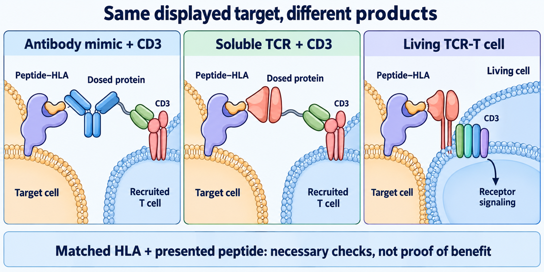
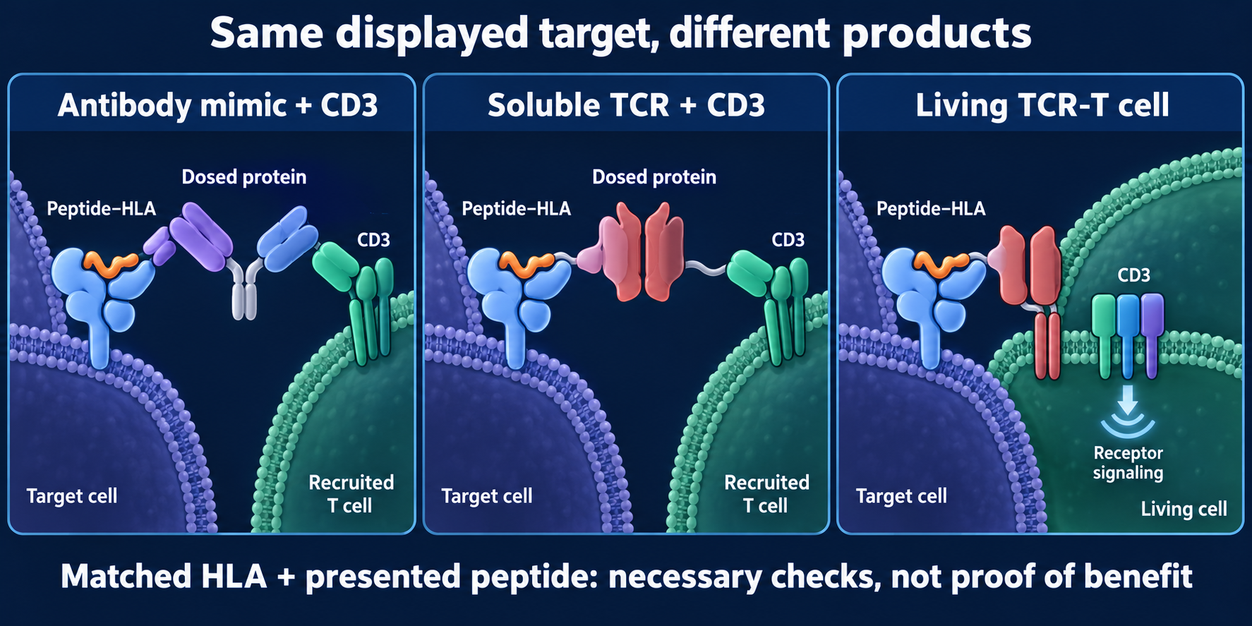
An antibody-derived T-cell receptor (TCR) mimic and an engineered soluble TCR can each be joined to a CD3-binding arm in a dosed protein. A living TCR-T cell instead carries a receptor with associated signaling. All require the specified peptide–human leukocyte antigen (HLA) target; matching recognition requirements does not establish activity, safety or clinical benefit. Shapes and sizes are schematic.
For a peptide–HLA/CD3 protein, the recruited T cell need not have a native TCR specific for that peptide. The target cell still needs the recognized complex. Bypassing the effector's native specificity does not bypass target HLA restriction or the need for functional T cells.
Why it matters in cancer
Intracellular proteins offer potential target fragments, but recognition requires actual display. Healthy cells may also present the intended complex; unintended peptide recognition is another safety question. A higher binding affinity or a matched allele cannot establish a safety window or clinical benefit.
A scoped clinical example
Tebentafusp is a soluble gp100 peptide–HLA-A02:01 TCR/CD3 protein. Its August 2026 label specifies HLA-A02:01-positive adults with unresectable or metastatic uveal melanoma. This illustrates an allele-restricted product and defined clinical context, not transferable breast-cancer benefit.
How it is measured
| Observation card | Record and interpret |
|---|---|
| Inputs | Actual protein/cellular product, HLA-typed target cells and relevant T-cell source; culture and binding aliquots are consumed |
| Target evidence | Exact peptide sequence, allele, surface-complex detection and recovery limits; source-protein or ribonucleic acid (RNA) abundance is different |
| Function | Concentration, duration, effector-to-target ratio, target-cell death and activation separately; percentages require a defined cell denominator |
| Controls and limits | Wrong-allele, peptide/target-negative, no-effector and relevant healthy-cell controls; peptide loading can bypass natural processing; no universal display cutoff or model-to-clinical conversion |
Common confusions
- TCR mimic does not mean engineered TCR. The recognizing scaffold is antibody-derived.
- Source protein does not mean presented target. The peptide–HLA complex needs its own evidence.
- Soluble receptor does not mean living cells. Product persistence and control differ.
- Compatible HLA is necessary for a specified route, not sufficient for activity. Display, exposure and immune function remain to test.
Try it
Fictional Cells A and B make source protein Q. A displays the recognized Q–HLA complex; B has a different HLA allele. A third cell has the compatible allele, but no display assay. Can all three be assumed to respond to the same peptide–HLA/CD3 protein?
Answer: No. A has target-display evidence, but still needs functional and safety testing. B lacks the specified recognition frame. The third has genotype evidence without display evidence. Protein Q abundance supplies neither missing result.
Explain it back
“The recognition route is shared, but the administered products differ because ___.”
One answer: an antibody scaffold, a soluble TCR/CD3 protein and receptor-bearing living T cells connect recognition to action differently.
Takeaway
Keep the exact peptide–HLA target, recognition scaffold and administered product visible in every claim.
Related concepts
Sources and scope
Source check: October 10, 2026. Mechanism studies are limited to their constructs/models; the label example is product-, allele- and disease-specific. Practice is fictional; expert and learner review remain pending.
- Dao et al., 2015 — antibody-derived peptide–HLA/CD3 construct in preclinical studies.
- Liddy et al., 2012 — engineered soluble TCR/CD3 constructs in experimental models.
- Tebentafusp label, August 2026, sections 1, 2.1 and 11–12.1 — administered protein, exact target and defined indication.
- Morgan et al., 2006 — transfer of receptor-engineered lymphocytes in a defined melanoma study, not evidence for all cell/protein formats.