Immunopeptidome: the biological display repertoire
In one sentence
The immunopeptidome is the repertoire of peptides associated with human leukocyte antigen molecules in a defined cell population and biological context.
The intuition
Think of a changing exhibition. The collection is what is displayed in that particular place and time. A visitor's notebook records only what they noticed. Likewise, a cell's peptide repertoire and a laboratory's recovered list are related but different objects.
The analogy has limits: peptides are generated, loaded and removed continuously, and laboratory recovery has chemical and computational biases. A notebook's omissions cannot tell us how many exhibits were truly absent.
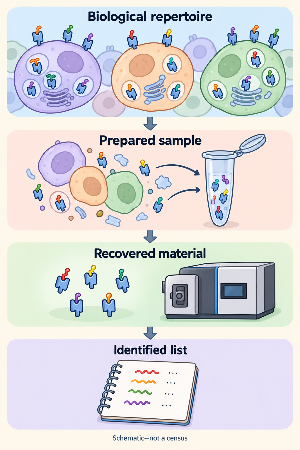
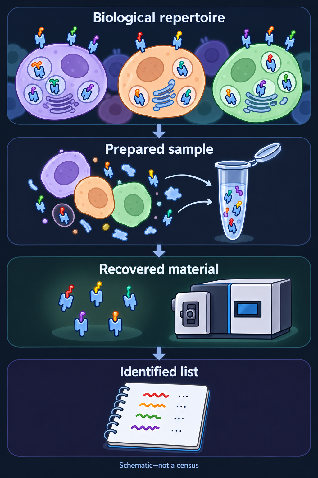
Preparation, recovery and sequence identification shape the list; recovered complexes are processed to release peptides for identification. Whole-cell extraction can include intracellular and surface complexes, and mixed tissue leaves each peptide's cell of origin open. Icons have no numerical meaning. A missing entry does not prove biological absence; a listed peptide does not establish recognition or benefit.
How it works
Human leukocyte antigen (HLA) molecules carry short protein fragments, or peptides. Different alleles favor different sequences. Antigen-processing machinery, source-protein supply, loading and turnover shape which complexes exist.
Class I and class II have distinct common processing routes. A class-I repertoire is not a census of class-II display. Nor is it a list of all cellular proteins: a protein can be abundant while a particular derived peptide is poorly generated, loaded or retained.
The repertoire depends on cell identity and state. In ovarian- and lung-cancer cell-line experiments, inflammatory signals changed HLA-associated peptide patterns. That demonstrates context-dependent remodeling; it does not mean inflammation always adds useful tumor targets. One specimen's repertoire need not represent another site, time or treatment condition.
For recognition at a living cell's surface, the relevant object is the surface peptide–HLA complex. An assay that captures HLA from whole-cell lysate can also recover intracellular complexes. The biological claim must therefore say which compartment was examined.
Why it matters in cancer
A vaccine board starts with candidate sequences. The immunopeptidome concerns the subsequent presentation layer. A T-cell receptor must still recognize a suitable peptide–HLA pair; presentation alone does not establish activation, killing, safety or patient benefit.
Mixed tumor tissue adds a source question. Cancer, immune and stromal cells can contribute peptides. An identified target in that mixture is not automatically cancer-cell-specific or present on every cancer cell.
How it is measured
Immunopeptidomics captures HLA-associated material and identifies recovered peptides, commonly by liquid chromatography and tandem mass spectrometry. The method page owns the input, capture and identification details. Confidence matters too: a reported sequence can be incorrectly assigned.
| Field | Keep attached to the repertoire claim |
|---|---|
| Biological scope | Cell population, specimen site, time, state and HLA class/alleles |
| Readout | Identified peptide sequences and their evidence; unique counts depend on the workflow |
| Abundance | Relative instrument intensity is not automatically surface copies per cell |
| Coverage | Capture reagent, recovery, search database and identification threshold |
| Material and cost | Method-specific consumed tissue/cells and analysis requirements |
| Validation limit | Non-detection is not universal absence; recovery is not recognition or clinical utility |
Common confusions
- Biological repertoire versus recovered subset: the reported list is shaped by sampling and measurement.
- Protein expression versus peptide display: the source and the displayed fragment have different dependencies.
- More identified peptides versus better immunity: a larger list can reflect recovery or ordinary self peptides.
- Mixed specimen versus cancer-cell surface: cell origin and compartment need evidence.
Try it
Two fictional laboratories analyze comparable portions of one specimen. Method A identifies 800 peptides and method B identifies 1,200. Has B shown that those cells biologically displayed 50% more peptides?
Answer: No. The counts describe identified sets under two workflows. Capture, sensitivity and analysis could account for the difference. These invented numbers supply no recovery fraction or clinical threshold.
Explain it back
“The immunopeptidome is ___; an immunopeptidomics list is ___.” One answer: “a biological repertoire in context; a method-dependent set recovered and identified from a sample.”
Takeaway
Name the biological set and the measurement's subset separately.
Related concepts
Sources and scope
Source check: October 10, 2026. General repertoire biology and a fictional comparison; expert and learner review remain pending.
- Sarkizova et al., 2020, allele-dependent peptide repertoires in monoallelic cell lines and tumor-line validation.
- Chong et al., 2018, ovarian-cancer cell experiments and interferon-associated repertoire changes.
- Javitt et al., 2019, inflammatory signals, HLA-B expression and peptide patterns in lung-cancer cell experiments.
- Purcell et al., 2019, HLA-associated peptide isolation and identification protocol.
- Wilhelm et al., 2021, analytical sensitivity and identification limits.