Read each label as an answer to a question
A cancer report gives several answers about one specimen. Keep each answer attached to its question before combining them into a picture.
Before you start: Tumor heterogeneity explains why different cells and regions can differ. This matters whenever a small specimen is used to describe a larger tumor.
Where this step sits
This is step 1 of Start here. First read the labels. Next follow an immune response. Then connect a treatment claim with its evidence.
Begin with the specimen
Cancer develops when cells acquire changes that let them grow and survive outside normal controls. Some can invade nearby tissue and spread. A tumor can also contain immune cells, support cells and treated or damaged tissue. The name of a tumor does not mean every cell in a sample is a cancer cell.
Ask where the specimen came from and when it was collected. A biopsy before treatment and a surgical specimen after treatment describe different moments. A report date tells you when a report was issued; it does not replace the collection date. An addendum may answer a new question about the earlier specimen.
A pathologist examines the tissue and integrates the relevant tests. The final interpretation deserves priority over a striking word in the microscopic description. If a field is pending or qualified, keep that qualification beside it.
Give each label a job
| Question | A label or result you may see | What it does not answer by itself |
|---|---|---|
| What tissue pattern is present? | Histologic type and invasive versus in situ disease | The full extent throughout the body |
| What does the cancer look like microscopically? | Histologic grade | The stage or an exact growth rate |
| What receptor features were found? | Estrogen receptor, progesterone receptor and HER2 (human epidermal growth factor receptor 2) tests | Every possible treatment target |
| What expression pattern was assigned? | Intrinsic molecular subtype | A replacement for the receptor test |
| What extent is established? | Tumor, node and metastasis (TNM) staging | A certain personal outcome |
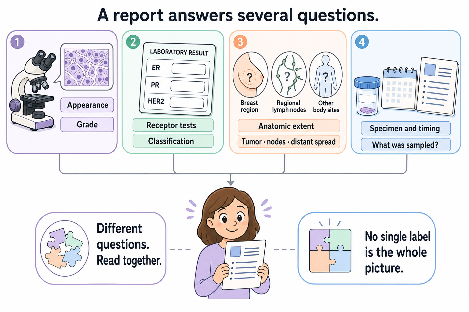
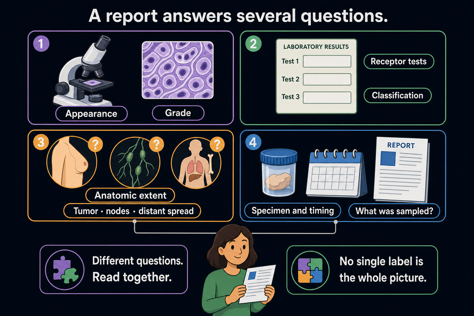
Each panel asks a different question. Some breast prognostic staging systems combine anatomic extent with grade and biomarkers. This cartoon organizes those observations; it is not a patient report, a stage assignment or a forecast.
HER2 means human epidermal growth factor receptor 2. It is a protein and its associated testing category, rather than a name for all growth signals. Estrogen receptor and progesterone receptor are separate hormone receptors. Their reports help define hormone-receptor status.
Triple-negative breast cancer (TNBC) is a receptor-based category. The name does not mean the cancer has no proteins, no molecular differences or no treatment options. Different tumors within the category can have different biology.
A worked report: two descriptions can both be true
Imagine a fictional biopsy describes a high-grade invasive breast cancer with triple-negative receptor classification. Imaging also identifies a breast lesion and a suspicious regional node. The node has not yet been sampled.
“High grade” describes microscopic features. It does not establish distant spread. “Triple-negative” describes the tested receptor category. It does not establish a particular ribonucleic acid (RNA) expression subtype. The suspicious node is a clinical finding that needs its own interpretation; it is not automatically a signed positive-node pathology result.
Now imagine a later surgical report finds sparse residual invasive cancer in a treated tumor bed. That later finding does not rewrite the earlier biopsy into a post-treatment measurement. You need both dates and settings to understand the change.
What can go wrong at this step
- Treating grade as stage merges appearance with extent. Some breast prognostic staging systems also incorporate grade and biomarkers; read the named staging system.
- Treating receptor status as a whole molecular portrait leaves out other measured features.
- Treating a sample result as a survey of every cell ignores sampling and heterogeneity.
- Treating an unsigned estimate or pending result as final removes information the report still needs.
Try it
A fictional report says “grade 3.” A friend reads this as “stage III” and assumes distant spread. Which two steps in that reasoning fail?
Answer: Grade 3 is a microscopic grade, not a stage assignment. Stage III breast cancer also does not mean distant metastasis; distant spread is a separate part of staging. The actual clinical or pathologic staging assessment is needed.
Explain it back
“This label describes ___, in a specimen collected ___; it does not by itself tell me ___.”
One possible answer: “Histologic grade describes microscopic features, in a specimen collected before treatment; it does not by itself tell me the disease's extent.”
Takeaway
Keep the specimen, date, method and question beside every cancer label.
Next: Follow the route to immune recognition.
Sources and scope
General report-reading education, source-checked October 9, 2026; expert and learner review pending. The report is fictional and is not a staging calculation.
- NCI: What is cancer?, for altered cell behavior and tumor diversity.
- NCI: Pathology reports, for specimen, description and final diagnosis.
- NCI: Breast cancer stages, for staging dimensions. The linked receptor and grade concepts provide their testing sources.